
Multi-site neuroimaging studies offer unprecedented opportunities to investigate aging and neurodegenerative disease in large, diverse populations. However, differences among sites—including scanner hardware, acquisition protocols, software versions, participant populations, and image-processing pipelines—can introduce variability that complicates the interpretation, integration, and reproducibility of neuroimaging results.
This two-day symposium and workshop will examine best practices for designing, conducting, and analyzing multi-site neuroimaging studies of aging and neurodegeneration. Speakers will consider challenges that arise throughout the research lifecycle, from initial study planning and protocol development to scanner upgrades, unexpected acquisition changes, data processing, statistical analysis, and clinical translation. Particular attention will be given to distinguishing meaningful biological variability from variability introduced by sites, scanners, and analytical methods.
The program will introduce prospective and retrospective approaches for improving the comparability of data collected across institutions, scanner platforms, and software environments. Examples will span diffusion MRI, structural MRI and measures of vascular brain injury, magnetic resonance spectroscopy, high-field vascular imaging, and amyloid positron emission tomography. Speakers will also discuss statistical and AI-based harmonization methods, including their assumptions, limitations, and effects on downstream analyses.
The event will further highlight the value of community-developed standards and harmonized analysis protocols, including the STRIVE standards for vascular brain injury and the Hippocampal Subfields Group harmonized segmentation protocol. Through lectures, panel discussions, and practical workshops, participants will explore how harmonization, interoperability, open science, and emerging AI tools can support more reliable, scalable, and clinically meaningful neuroimaging research.
This event is part of the Advancing Aging and Neurodegeneration Research through Data Science Emergent Data Science Program.
By the end of this event, participants will be able to:
| September 28, 2026 | |||||||||||||||||||||||||||||||||||||||||||||||||||||||||||||||||||||||||||||||||||||||||||||||||||
|---|---|---|---|---|---|---|---|---|---|---|---|---|---|---|---|---|---|---|---|---|---|---|---|---|---|---|---|---|---|---|---|---|---|---|---|---|---|---|---|---|---|---|---|---|---|---|---|---|---|---|---|---|---|---|---|---|---|---|---|---|---|---|---|---|---|---|---|---|---|---|---|---|---|---|---|---|---|---|---|---|---|---|---|---|---|---|---|---|---|---|---|---|---|---|---|---|---|---|---|
|
8:55-9:00 am |
Opening remarks Rosanna Olsen Senior Scientist, Rotman Research Institute, Baycrest; and University of Toronto |
||||||||||||||||||||||||||||||||||||||||||||||||||||||||||||||||||||||||||||||||||||||||||||||||||
|
9:00-9:30 am |
From Hazard to Harmony: An Introduction to Harmonization Best Practices
Cheryl McCreary, Cumming School of Medicine, Department of Clinical Neurosciences, and Department of Radiology, University of Calgary |
||||||||||||||||||||||||||||||||||||||||||||||||||||||||||||||||||||||||||||||||||||||||||||||||||
|
9:30-10:30 am |
Inter-scanner MRI data harmonization – How critical is it? Yogesh Rathi, Office for Graduate Education PhD Programs, Harvard Medical School, Harvard University |
||||||||||||||||||||||||||||||||||||||||||||||||||||||||||||||||||||||||||||||||||||||||||||||||||
|
10:30-11:00 am |
Break
|
||||||||||||||||||||||||||||||||||||||||||||||||||||||||||||||||||||||||||||||||||||||||||||||||||
|
11:00-12:00 |
Evidence-Based Delphi Procedure with International Scholars to Develop the Hippocampal Subfield Group Harmonized Segmentation Protocol Ana Daugherty, Department of Psychology and the Institute of Gerontology, Wayne State University |
||||||||||||||||||||||||||||||||||||||||||||||||||||||||||||||||||||||||||||||||||||||||||||||||||
|
12:00-1:15 pm |
Networking and lunch |
||||||||||||||||||||||||||||||||||||||||||||||||||||||||||||||||||||||||||||||||||||||||||||||||||
|
1:15-2:15 pm |
Harmonization of Amyloid-PET Quantification Using the Centiloid Scale: Concept, Validation, and Clinical Context of Use Renaud La Joie, Memory and Aging Center; Department of Neurology, University of California, San Francisco |
||||||||||||||||||||||||||||||||||||||||||||||||||||||||||||||||||||||||||||||||||||||||||||||||||
|
2:15-3:15 pm |
Opportunities for MR spectroscopy in aging Ashley Harris, Cumming School of Medicine, Department of Radiology, University of Calgary |
||||||||||||||||||||||||||||||||||||||||||||||||||||||||||||||||||||||||||||||||||||||||||||||||||
|
3:15-3:30 pm |
Break |
||||||||||||||||||||||||||||||||||||||||||||||||||||||||||||||||||||||||||||||||||||||||||||||||||
|
3:30-4:30 pm |
Zooming In, Scaling Out: High-Resolution Vascular Imaging for Brain Aging and Neurodegeneration Saskia Bollmann, School of Electrical Engineering and Computer Science, The University of Queensland |
||||||||||||||||||||||||||||||||||||||||||||||||||||||||||||||||||||||||||||||||||||||||||||||||||
|
4:30-5:15 |
Panel discussion
|
||||||||||||||||||||||||||||||||||||||||||||||||||||||||||||||||||||||||||||||||||||||||||||||||||
|
5:15-5:20 |
Closing remarks |
||||||||||||||||||||||||||||||||||||||||||||||||||||||||||||||||||||||||||||||||||||||||||||||||||
| September 29, 2026 | |||||||||||||||||||||||||||||||||||||||||||||||||||||||||||||||||||||||||||||||||||||||||||||||||||
|---|---|---|---|---|---|---|---|---|---|---|---|---|---|---|---|---|---|---|---|---|---|---|---|---|---|---|---|---|---|---|---|---|---|---|---|---|---|---|---|---|---|---|---|---|---|---|---|---|---|---|---|---|---|---|---|---|---|---|---|---|---|---|---|---|---|---|---|---|---|---|---|---|---|---|---|---|---|---|---|---|---|---|---|---|---|---|---|---|---|---|---|---|---|---|---|---|---|---|---|
|
8:55-9:00 am |
Opening remarks Rosanna Olsen Senior Scientist, Rotman Research Institute, Baycrest; and University of Toronto |
||||||||||||||||||||||||||||||||||||||||||||||||||||||||||||||||||||||||||||||||||||||||||||||||||
|
9:00-10:00 am |
Harmonizing Measurement of Vascular Brain Lesions
Eric Smith, Cumming School of Medicine, Department of Clinical Neurosciences, University of Calgary |
||||||||||||||||||||||||||||||||||||||||||||||||||||||||||||||||||||||||||||||||||||||||||||||||||
|
10:00-11:00 am |
High-Resolution Imaging of Healthy Ageing and Disease at 3T and 7T: Accessibility and Clinical Opportunities Sriranga Kashyap, Sunnybrook Research Institute |
||||||||||||||||||||||||||||||||||||||||||||||||||||||||||||||||||||||||||||||||||||||||||||||||||
|
11:00-11:30 am |
Break
|
||||||||||||||||||||||||||||||||||||||||||||||||||||||||||||||||||||||||||||||||||||||||||||||||||
|
11:30 am-12:30 pm |
Beyond ComBat: Next-generation harmonization methods for multi-centre neuroimaging studies Russell Taki Shinohara, Department of Biostatistics, Epidemiology & Informatics, University of Pennsylvania |
||||||||||||||||||||||||||||||||||||||||||||||||||||||||||||||||||||||||||||||||||||||||||||||||||
|
12:30-1:30 pm |
Networking and lunch |
||||||||||||||||||||||||||||||||||||||||||||||||||||||||||||||||||||||||||||||||||||||||||||||||||
|
1:20-2:30 pm |
From Heterogeneous Data to Biological Insight: Multimodal Data Science for Brain Aging AmanPreet Badhwar, Department of Pharmacology and Physiology and the Institute of Biomedical Engineering, Institut universitaire de gériatrie de Montréal (CRIUGM), Université de Montréal |
||||||||||||||||||||||||||||||||||||||||||||||||||||||||||||||||||||||||||||||||||||||||||||||||||
|
2:30-2:45 pm |
Break
|
||||||||||||||||||||||||||||||||||||||||||||||||||||||||||||||||||||||||||||||||||||||||||||||||||
|
2:45-4:15 pm |
Workshop 1, Room 10031 Neurodesk: An application promoting reliability, interoperability, and the democratization of data analysis Steffen Bollmann, School of Electrical Engineering and Computer Science, Queensland Digital Health Centre, University of Queensland Workshop 2, Room 10032 Hands on Segmentation Training Ana Daugherty, Department of Psychology and the Institute of Gerontology, Wayne State University Workshop 3, Mini Auditorium, Mezzanine level Agentic analysis of fMRI data, considerations for data privacy Bradley Buchsbaum, Rotman Research Institute (RRI), Baycrest, Department of Psychology, Faculty of Arts & Science, University of Toronto |
||||||||||||||||||||||||||||||||||||||||||||||||||||||||||||||||||||||||||||||||||||||||||||||||||

AmanPreet Badhwar
Department of Pharmacology and Physiology and the Institute of Biomedical Engineering, Research Centre of the Institut universitaire de gériatrie de Montréal (CRIUGM), Université de Montréal
Prof. Badhwar’s research seeks to understand the complex interplay of vascular, metabolic, genetic, and environmental factors that contribute to brain aging and dementia, particularly Alzheimer’s disease and vascular cognitive impairment. She employs advanced techniques in neuroimaging (7T MRI, functional connectivity), bioinformatics, machine learning, and extracellular vesicle biology to identify early predictive and subtype-specific biomarkers of dementia. Her current projects focus on investigating blood- and protein-based biomarkers of MRI-detected cerebrovascular injury, studying the impact of biological sex, gender, and menopause on cognitive aging and neurodegeneration, co-developing educational tools for individuals living with cognitive decline and their caregivers and working to improve equity, diversity, and inclusion in aging and brain health research cohorts.
Saskia Bollmann
School of Electrical Engineering and Computer Science, The University of Queensland
Prof. Bollmann obtained her M.Sc. ETH in Biomedical Engineering from the Swiss Federal Institute of Technology Zurich in 2013, where she utilized concurrent magnetic field monitoring to improve image quality and temporal stability of functional magnetic resonance imaging (fMRI). She then moved to Brisbane in 2014 to pursue a PhD at the Centre for Advanced Imaging, The University of Queensland, under the supervision of Prof. Markus Barth. She investigated contrast mechanisms and signal properties of fMRI focusing on ultra-high field applications. In 2019, she joined A/Prof. Jonathan Polimeni at the Martinos Center for Biomedical Engineering, Massachusetts General Hospital, where she employed ultra-high field MRI to image the blood vessels of the human brain and better understand their contribution to the fMRI signal. She returned to Brisbane in 2020 and is currently a Research Fellow at the School of Information Technology and Electrical Engineering, Faculty of Engineering, Architecture and Information Technology. She is developing new contrast mechanisms for functional MRI and investigating the effect of the vasculature on the fMRI signal.
Steffen Bollmann
School of Electrical Engineering and Computer Science, Queensland Digital Health Centre, University of Queensland
Prof. Bollmann’s research expertise is in quantitative susceptibility mapping, image segmentation and software applications to help researchers and clinicians access data and algorithms. He completed his PhD on multimodal imaging at the University Children’s Hospital and Swiss Federal Institute of Technology (ETH) Zurich, Switzerland. In 2014 he joined the Centre for Advanced Imaging at UQ as a National Imaging Facility Fellow, where he pioneered the application of deep learning methods for quantitative imaging techniques, in particular Quantitative Susceptibility Mapping. In 2019, he joined the Siemens Healthineers collaborations team at the MGH Martinos Center in Boston on a one-year industry exchange where he worked on the translation of fast imaging techniques into clinical applications

Bradley Buchsbaum
Rotman Research Institute (RRI), Baycrest, Department of Psychology, Faculty of Arts & Science, University of Toronto
Prof. Buchsbaum aims to understand how the varieties of conscious memory experience may be understood at the level of brain networks and activation patterns. He uses functional magnetic resonance imaging (fMRI) to capture and “decode” the patterns of neural activity that give rise to memory experiences. Using statistical techniques and machine learning methods, He is developing ways of “reading out” the content, precision, and quality of a person’s memory from the functional images that we acquire during the act of remembering.
Ana Daugherty
Department of Psychology and the Institute of Gerontology, Wayne State University
Prof. Daugherty is a cognitive neuroscientist with a specialty in aging, Alzheimer’s disease and related dementia. Her research characterizes individual differences in aging across the adult lifespan to identify both risk and protective factors that modify changes in the brain, and related changes in thinking and memory functions. The long-term goal of her work is to reduce disparities in late-life cognitive health. She directs the Detroit Aging Brain Study: a community-partnered longitudinal study going on over 22 years in the Metro Detroit area to study changes in brain structure and function across the healthy, adult lifespan.
Ashley Harris
Cumming School of Medicine, Department of Radiology, University of Calgary
Prof. Harris’s research interests involve the development and use of advanced imaging to study the brain in humans. This includes both health brain function and clinical disorders including developmental disorders, brain injury, migraine and chronic pain. In particular she develops and uses advanced spectroscopy, an approach to study brain chemistry in addition to other using other MR modalities such as functional and structural imaging.

Sriranga Kashyap
Sunnybrook Research Institute
Dr Kashyap is an interdisciplinary neuroscientist with over 12 years of ultra-high-field (7 T) MRI experience and a background spanning biomedical engineering, MR physics, neurophysiology, and cognitive and clinical neurosciences. Working closely with neurologists, neuroradiologists, and neurosurgeons at Toronto Western and Sunnybrook Hospitals, he serves as co-PI on three ongoing multi-year clinical MRI studies spanning neuromodulation, movement disorders, and neurodegeneration.
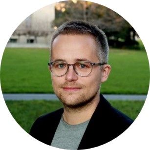
Renaud La Joie
Memory and Aging Center; Department of Neurology, University of California, San Francisco
Prof. Le Joie’s research contributions primarily focus on medical and neuroscience fields, with a strong emphasis on neurodegenerative disorders. The main areas of study include medicine and neuroscience, while subfields encompass psychiatry and mental health, physiology, cognitive neuroscience, radiology, nuclear medicine and imaging, and neurology.
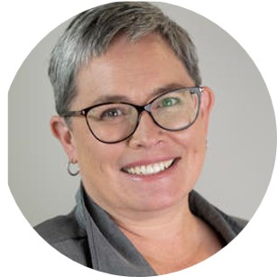
Cheryl McCreary
Cumming School of Medicine, Department of Clinical Neurosciences, and Department of Radiology, University of Calgary
Prof. McCreary had a diverse background, applying imaging techniques to cells, tissues, animal models, and people. These techniques included fluorescence spectroscopy to track calcium signaling in lymphocytes, in vivo proton and phosphorus magnetic resonance (MR) spectroscopy of the heart and brain, MR imaging of white matter to evaluate myelin in a murine model of multiple sclerosis, and MR imaging biomarkers of neurodegeneration associated with cerebral small vessel disease and aging. Since 2009, she worked with Drs. Eric Smith and Richard Frayne as an imaging research scientist and MR research manager, developing novel, non-invasive MRI markers of small vessel disease and cognitive impairment in aging. This included developing, optimizing, and troubleshooting MR protocols for local, national, and international research studies and clinical trials; managing image data transfer, curation, and quality review; and image processing for quantitative metrics.
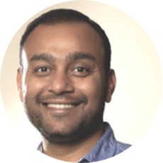
Yogesh Rathi
Office for Graduate Education PhD Programs, Harvard Medical School, Harvard University
Prof. Rathi’s research interests lie in developing smart imaging techniques to understand brain structure and function. He has developed several compressed sensing algorithms for fast imaging of diffusion MRI — a technology that is now being used to obtain advanced MRI scans of children with ADHD (Attention-deficit Hyperactivity Disorder). His current research focus is on 1). Ultra-high resolution diffusion imaging combining acquisition and reconstruction, 2). estimating tissue microstructure from biophysical and stochastic models of diffusion, and 3). time-series analysis for understanding functional connectivity using fMRI, EEG or MEG data. His clinical research focus includes using and developing sophisticated tractography algorithms for precise targeting of deep-brain stimulation (DBS) and transcranial magnetic stimulation (TMS) in obsessive compulsive disorder (OCD), Parkinson’s and major depressive disorder (MDD). His broad research focus is in the areas of signal and image-processing, statistics, control theory, machine learning, computer vision and related applications to solve inverse problems in medical imaging.
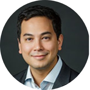
Russell Taki Shinohara
Department of Biostatistics, Epidemiology & Informatics, University of Pennsylvania
Prof. Shinohara’s methodological research centers on the assessment of structural and functional changes in the brain throughout the lifespan and in neurological, psychiatric, and developmental disorders. He is interested in describing complex processes and studying questions that have direct impacts on human health. He pursues broad collaborative interests in medicine and public health, with particular emphasis in quantitative biomedical imaging and biomarkers.
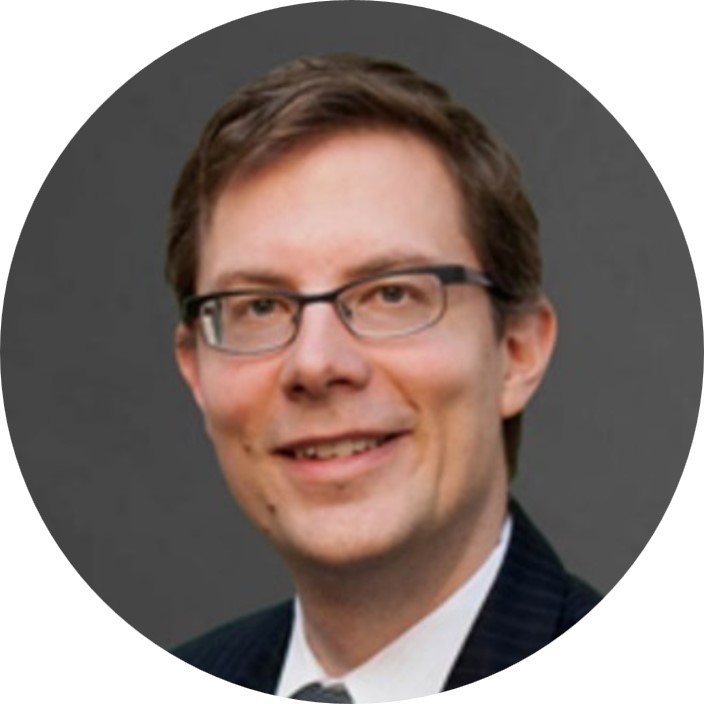
Eric Smith
Cumming School of Medicine, Department of Clinical Neurosciences, University of Calgary
Prof. Smith directs the Clinical and Research Fellowship program in Cognitive Neurosciences. Dr. Smith graduated from McGill University, trained in Neurology in teaching hospitals of Harvard Medical School, and was Assistant Professor of Neurology at Harvard University before being recruited to Calgary in 2008. He Co-Chairs the Canadian Stroke Best Practices Advisory Committee of the Heart and Stroke Foundation, and is a member of the Executive Committee of the Canadian Consensus Conference on the Diagnosis and Treatment of Dementia. Dr. Smith’s research team investigates how cerebral small vessel diseases cause cognitive impairment and dementia.
November 10 – 11, 2023
In-person only
10th floor seminar room
700 University Avenue,
Toronto, ON
In person
Data Sciences Institute,
Seminar room
10th floor,
700 University Avenue